What Causes Abnormal Ecg
ECG Yellow Belt online course. Atrial depolarization is in a different direction.

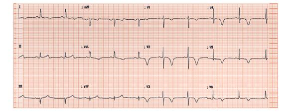
Normal 12 Lead Ecg Complexes Icu Nursing Normal Ecg Cardiology Nursing
ECG Blue Belt online course.

What causes abnormal ecg. An abnormal ECG can signal that one or more aspects of the hearts walls are larger than another meaning that the heart is working harder than normal to pump blood. An electrocardiogram is a test to measure how the electricity in a persons heart is functioning. While an abnormal EKG is helpful in making the diagnosis in the early stages of inflammation the EKG may be normal.
Wiesbauer F Kühn P. The goal of therapy is to treat the. Become an ECG expert.
Abnormal Patterns Possible Causes. Defects or abnormalities in the hearts shape and size. The electrocardiogram EKG or ECG shows electrical activity of the heart.
If the ECG is abnormal at rest the patient should undergo a thallium stress test or exercise echocardiography. The heart is a muscle that contracts and relaxes to pump blood around the body. An abnormal ECG can also tell your healthcare professional if you have any of these.
An abnormal heart rhythm is when your heart beats too fast too slow or irregularly. Abnormal heart rhythms can also result and ECG findings of a short QT interval suggest hypercalcaemia. RAE is suggested by an ECG which has a pronounced notch in the P wave.
The slow spread of the impulse will result in a slow and abnormal activation of the right ventricle which yields a bizarre and prolonged QRS complex on ECG. Since left ventricular depolarization is abnormal the repolarization will also be abnormal and secondary ST-T changes are always present. Within the heart is a complex system of valves nodes and chambers that.
Electrolytes are electricity-conducting particles in the. This causes a wide S-wave in V1V2 it is referred to as QS complex if the r-wave is absent and broad and clumsy R-wave in V5V6. There can be a number of causes for a borderline electrocardiogram ECG a study of electrical activity in the heart.
Learn to diagnose any rhythm problem. But if atrial fibrillation is present a P wave would not be present. Wiesbauer F Kühn P.
In pericarditis there are hallmark changes that are seen and can help make the diagnosis. By positioning leads electrical sensing devices on the body in standardized locations health care professionals can learn information about many heart conditions by looking for characteristic patterns on the EKG. When an ECG is borderline it means that some anomalies are present and the doctor needs to evaluate the patient to.
An electrocardiogram also called an ECG or EKG is widely used as a screening test for right atrial enlargement. The heart is in an abnormal location or orientation within the chest dextrocardia. QRS Axis Determination First find the isoelectric lead if there is one.
Ie the lead with equal forces in the positive and negative direction. March 17 2022 An ECG procedure will result in a readout of electrical activity in the heart. Hypercalcaemia has also been known to cause an ECG finding mimicking hypothermia known as an Osborn wave.
This is also called an arrhythmia. The R-wave may be notched at the apex. Clinical ECG Interpretation ECG Waves.
Mattu A Tabas JA Brady WJ. Significant hypercalcaemia can cause ECG changes mimicking an acute myocardial infarction. The electrocardiogram ECG or EKG is a noninvasive test that is used to reflect underlying heart conditions by measuring the electrical activity of the heart.
An exercise ECG is important in identifying the presence of ischemic heart disease and the amount of myocardium at risk. QT prolongation may increase the risk of developing abnormal heart rhythms and may lead to sudden cardiac arrest. The electrocardiogram ECG or EKG is a diagnostic tool that is routinely used to assess the electrical and muscular functions of the heart.
Abnormal results can signify several issues. Pacemaker is no longer the SA node and is subsumed by another part of the heart. While it is a relatively simple test to perform the interpretation of the ECG tracing requires significant amounts of training.
Click to see causes of abnormal axis lesson 4. Greaten than 25mm in amplitude. Dr Smiths ECG blog.
The hallmark of right bundle branch block is QRS duration 012 seconds large R-wave in V1V2 and a. ECG and EKG are different abbreviations for the same test called an electrocardiogram. Mary McMahon Date.

Cardiac Syncope Ecg Interpretation Ecg Cases Em Cases
Learn About Left Bundle Branch Block Lbbb With Emphasis On Ecg Criteria Differential Diagnoses Cause Bundle Branch Block Cardiac Nursing Medical Knowledge

Ecg Hyperkalemia Hyperkalemia Nursing School Notes Cardiac Nursing

Abnormal Ecg Results Causes And Treatment Heart Care In Nairobi

Can T Miss Ecg Findings Cards For The Emergency Medicine Provider Cards Ecg Emergency Findings Medic Cardiac Nursing Icu Nursing Nursing School Survival

Pin On Cardiology Related Fields

S1q3t3 Ekg Classic Pattern In Pulmonary Embolism Example Pulmonary Embolism Ekg Pulmonology
Emotion And The Ecg Thoracic Key

5 Home Twitter Myocardial Infarction Medical Pictures Echocardiogram
Belum ada Komentar untuk "What Causes Abnormal Ecg"
Posting Komentar